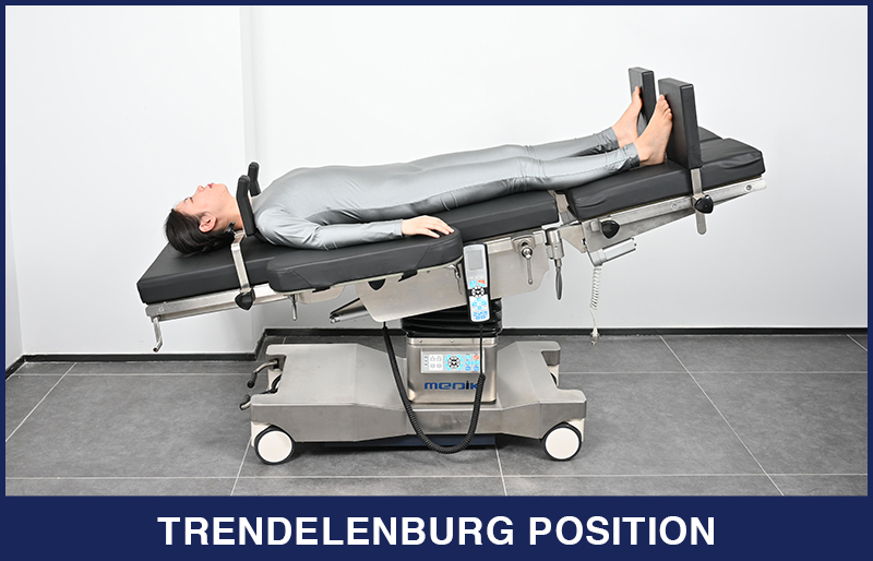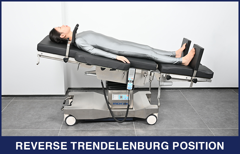

Positioning plays a crucial role in ensuring the safety of patients undergoing surgical procedures. The appropriate placement of the patient hinges on various factors such as the nature and duration of the procedure, the method of anesthesia, necessary medical devices, and other relevant considerations. While in Trendelenburg position, involves laying the patient supine on the surgical table with the head inclined downwards. By utilizing the force of gravity, this posture facilitates the surgical team's access to the abdominal organs. This is achieved by displacing surrounding organs within the abdominal cavity, thus granting unobstructed visibility and reach to the surgical site.
What Is The Trendelenburg Position?
The Trendelenburg position is a position for a patient on the operating table, most commonly used during lower abdominal surgeries and central venous catheter placement. In the Trendelenburg position, the body is laid supine, or flat on the back on a 15–30 degree incline with the feet elevated above the head.

When to use The Trendelenburg Position?
The Trendelenburg position is used in surgery, especially of the abdomen and genitourinary system. It allows better access to the pelvic organs as gravity pulls the intra-abdominal organs away from the pelvis. Evidence does not support its use in hypovolaemic shock, with concerns for negative effects on the lungs and brain.
Considerations For Trendelenburg Position
When arranging a patient in the Trendelenburg position, the medical team must carefully consider a range of factors, encompassing potential risks, susceptibility to injuries, and clinical directives regarding repositioning.
Brachial Plexus Neuropathy
– The proximity of the clavicle and the first rib poses a threat of compressing the subclavian vessels and the brachial plexus. The utilization of shoulder braces can contribute to brachial plexus neuropathy:
The force of gravity pushes the patient's weight onto the shoulder braces, causing the shoulders to give way under pressure, resulting in the body mass shifting between the braces.
Perioperative complications arising from extended Trendelenburg positioning
– Sustaining the Trendelenburg position for prolonged durations may increase the likelihood of various complications, such as ocular trauma due to heightened blood pressure in the head, including corneal abrasions, retinal detachments, and ischemic optic neuropathy. Furthermore, there is an associated risk of respiratory distress.
Clinical recommendations for repositioning patients
– Employing a steep Trendelenburg position raises the potential for intraoperative harm. To prevent injuries, proactive measures are crucial. Several strategies can be implemented, such as administering anesthesia through crystalloids to mitigate the risk of neural edema, surgeons opting for minimal pneumoperitoneum necessary for the surgery, and using the least degree of Trendelenburg required for the procedure. Vigilant monitoring of the patient's positioning should be carried out by the nursing team. For cases lasting over 4 hours, it has been suggested to conduct secondary timeouts to reevaluate patient positioning and make adjustments based on the patient's physiological and physical condition. Routinely, nursing staff should assess the safety of the patient's position every hour whenever feasible. Incorporating pauses for positioning assessments and periodically alleviating the Trendelenburg position can effectively diminish the likelihood of the most prevalent injuries sustained by patients in this stance.
Trendelenburg Position Vs. Reverse Trendelenburg Position
In the Trendelenburg position, the body is laid supine, or flat on the back on a 15–30 degree incline with the feet elevated above the head. The reverse Trendelenburg position, similarly, places the body supine on an incline but with the head now being elevated.
Reverse Trendelenburg position is used for laparoscopic surgeries including gallbladder, biliary tract, and stomach procedures, as well as head and neck surgeries.
Most recently, the Reverse Trendelenburg position has been used in minimally invasive glaucoma surgery, also known as MIGS. This position is commonly used for a superior sitting surgeon that uses a combination of downward patient tilt, of approximately 30 to 35 degrees, microscope tilt towards themselves at the same angle, and an intraoperative gonio lens or prisms that allows them to visualise the inferior trabecular meshwork.

Conclusion
Positioning a patient appropriately during a surgical intervention serves to minimize the risk of harm and enhance overall comfort. The Trendelenburg position, specifically, offers surgeons enhanced access to pelvic organs, proving beneficial for procedures such as colorectal, gynecological, and genitourinary surgeries. Nonetheless, akin to all surgical poses, a comprehensive risk assessment must precede the placement of a patient into the Trendelenburg position. It's crucial to recognize potential hazards associated with this posture. For instance, risks inherent in the Trendelenburg position comprise decreased lung capacity, reduced tidal volume, compromised pulmonary compliance, the pooling of venous blood towards the patient's head, and the potential for sliding and shearing forces. It's advisable to refrain from employing the Trendelenburg position in cases involving extremely obese patients.
*PLEASE NOTE: We are healthcare equipment suppliers, and therefore are not qualified to give any clinical or medical advice.*
References
1.Ostrow, CL (May 1997). "Use of the Trendelenburg position by critical care nurses: Trendelenburg survey". American Journal of Critical Care. 6 (3): 172–6.
2. Johnson, S; Henderson, SO (2004). Canadian Journal of Emergency Medicine. 6(1): 48–9. doi:10.1017/s1481803500008915. PMID 17433146.
3. Zillioux JM, Krupski TL. Patient positioning during minimally invasive surgery: what is current best practice? Robot Surg. 2017;4:69-76. Published 2017 Jul 14. doi:10.2147/RSRR.S115239